Top-tips: Chest X-Rays - Step 1, Technical quality of the radiographArticle 7 of 21
The key to reporting a chest X-ray is to understand the normal anatomy and to develop a systematic approach to your pattern of review. By following a stepwise approach the chance of missing abnormalities is minimized and will also provide a structured approach especially in an examination situation.
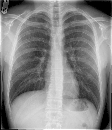
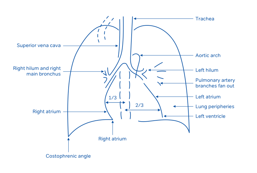
Figure 1: Normal chest X-ray
Reviewing a chest x-ray
Use the above film (fig 1) as a reference point for the rest of this part. In the OSCE Exam, the Chest XRay may be anonymised – you should say that the first thing you would look at is the patient identifier usually at the top corner. Say you need to confirm that the XRay belongs to the correct patient. Firstly, some technical details: Quickly look at the film to get some useful information about the patient at the same time as the quality of the film:
Step 1: Technical quality of the radiograph:
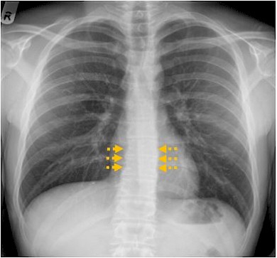
- Good penetration? You should just be able to see the lower thoracic vertebral bodies through the heart.
Figure 2: An adequately exposed film with the vertebral bodies (arrows) just visible through the heart.
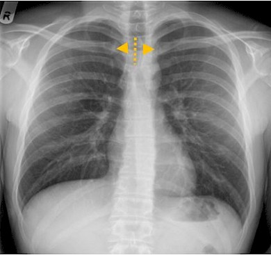
- Is the patient rotated? The spinous processes of the thoracic vertebrae should be midway between the medial ends of the clavicles.
Figure 3: The end of the clavicles (arrows) are equidistant from the midline indicating a non-rotated film.
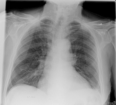
- PA or AP projection: Most chest x-ray films are taken posterior anterior (PA) - that is, the x-rays shoot through from the back of the patient to the x-ray plate in front of the patient. If the patient is too sick to stand up for this, an anterior posterior (AP) film will be done - that is, the x-rays shoot through from front to back. An AP film will always be labelled as ‘AP’, so if nothing is written on the film it is safe to assume it is PA. PA films are better, particularly because the heart is not as magnified as on an AP film, making it easier to comment on the heart size.
Tip: You can avoid the whole PA/AP debate by describing all chest X-rays as "frontal" - that is, you are looking at the patient straight on.
Figure 4: PA projection (the norm)
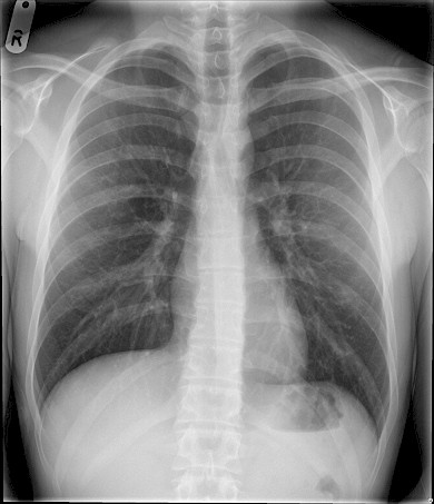
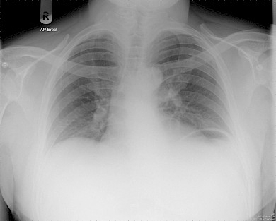
Figure 5: AP projection. As you will notice there is significant magnificent of the heart and mediastinal structures. This is usually labelled as 'AP film' on the radiograph.
- Good inspiration? It is easy to get tied up in knots over this - and sometimes not get any further. The diaphragms should lie at the level of the sixth ribs anteriorly. The right hemi-diaphragm is usually higher than the left because the liver pushes it up.
Figure 6: Good inspiratory film with 6 ribs seen anteriorly.
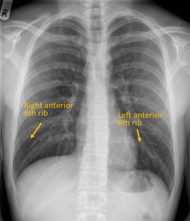
Figure 7: Chest X-ray taken during expiration