Top tips: Chest X-Rays - Step 3, Pleural spaceArticle 12 of 21
Normal pleura is invisible on a chest X-ray and becomes visible if there is an abnormality. Common pathologies related to pleura are pleural thickening, pleural effusion and pneumothorax.
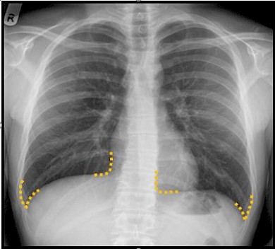
Figure 18: The costophrenic and cardiophrenic recess are indicated.
Pleural effusion:
- If the patient is erect there should be a fluid level and meniscus visible
- If the effusion is large the mediastinum will be shifted to the opposite side. Compare this with pure consolidation in which there is no change in volume of the hemi-thorax and therefore no mediastinal shift.
Figure 19: Blunting of costophrenic angles consistent pleural effsuion.
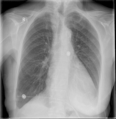
Figure 20: Massive left sided pleural effusion causing mediastinal and tracheal shift to the right.
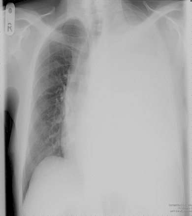
Figure 21: Large right hydro pneumothorax. Note the presence of air fluid level (represented by the straight line- arrow).
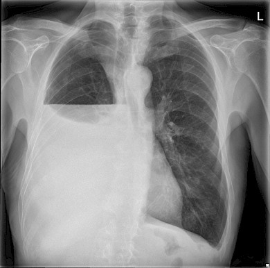
There is one caveat to bear in mind, which is that if collapse of the lung is accompanied by a pleural effusion the loss of volume (caused by the collapse) may be balanced out by the increase in volume of the hemi-thorax (caused by the effusion) and therefore it may seem as if the volume of the hemi-thorax overall is equivalent to the opposite side.
Pneumothorax:
Make sure you can see lung markings all the way to the edge of the chest wall. If the lung edge is visible with no lung marking beyond this then a pneumothorax should be suspected.
It is very easy to miss a pneumothorax. Watch out for the following signs:
- One half of the lung may seem blacker - that is, more radiolucent - than the other, which will be more radio-opaque or whiter. In particular, the area beyond the collapsed lung will be very radiolucent because there are no pulmonary vessel markings.
- You should be able to identify the edge of the collapsed lung.
Figure 22: Small right apical pneumothorax. Note the presence of a thin lung edge and absence of lung marking beyond this line.
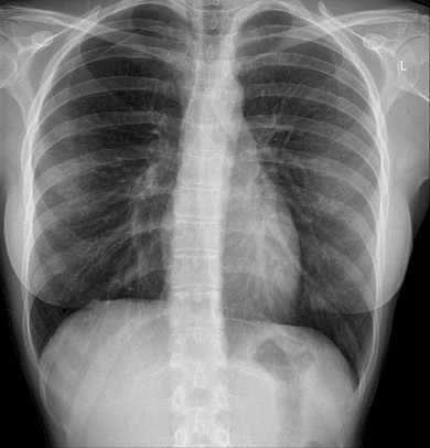
Having identified a pneumothorax you need to look for several more associated abnormalities:
- Most importantly - this is a pass or fail observation - is there evidence of a tension pneumothorax? This occurs when air can enter the pleural space (via a hole in the lung surface or the chest wall) but, because of a ball-valve effect, air cannot leave by the same route. So more and more air accumulates in the pleural space. This pushes the mediastinum over to the opposite (normal) side and eventually compresses the normal lung so that less inspiration occurs on the normal side, with compression on the heart and decreased venous return until finally the patient arrests. Always look for this and say: "There is no shift of the mediastinum and therefore no tension pneumothorax" or "There is shift of the mediastinum away from the side of the pneumothorax indicating a (right/left) tension pneumothorax. This is a medical emergency which I would treat immediately by inserting a large bore cannula into the (right/left) pleural space."
- The cause of the pneumothorax may be apparent - for example, fracture of the ribs.
- There may be associated surgical emphysema - that is, air in the soft tissues - and air in the mediastinum.
Figure 23: The lung edge is visible on the left consistent with a left pneumothorax.
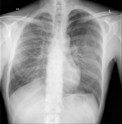
Figure 24: Large left sided tension pneumothorax causing mediastinal and tracheal shift to the right.
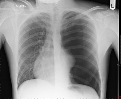
Pleural lesion:
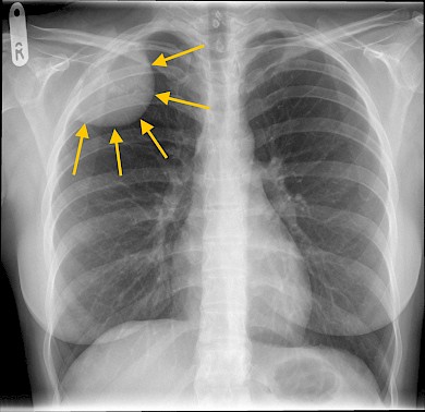
Figure 25: This is an example of a pleural based lesion. Notice the well-defined sharp medial margin and ill-defined lateral margin, a feature of pleural based lesions.