Top tips: Chest X-Rays - Step 3, Mediastinal contoursArticle 16 of 21
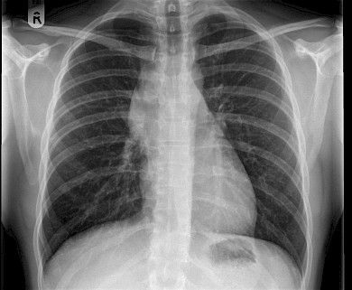
The mediastinum lies between the left and right lung. This comprises of heart and the great vessels (assigned as middle mediastinum), potential space in front of the heart (anterior mediastinum), behind the heart (posterior mediastinum) and above the heart (superior mediastinum). Important structures contributing to mediastinal borders are the aortic knuckle, the aorto-pulmonary window, and the right para-tracheal stripe.
The most common sign of mediastinal disease is mediastinal widening. This can result from masses in the anterior, middle and posterior mediastinum.
Figure 36: Important landmarks on a chest x ray; aortic knuckle, AP window and right paratracheal stripe.
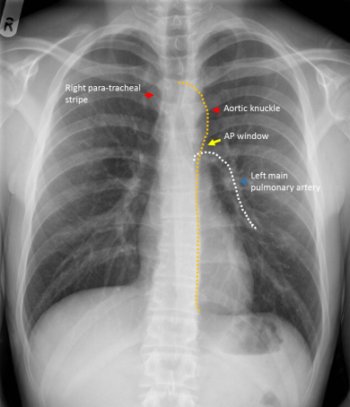
Figure 37: Notice the widening of the right paratracheal stripe and also soft tissue left mediastinum and AP window. The differential diagnosis would include lymphadenopathy.
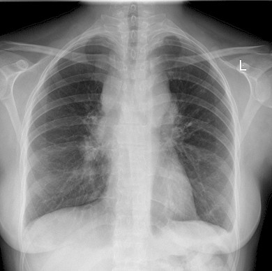
Figure 38: Widening of right paratracheal stripe in a patient with lymphoma