Top tips: Chest X-Rays - Step 3, HilumArticle 14 of 21
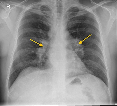
The hilum is an important landmark on a chest X-ray. The hilum is made of major bronchi and pulmonary vessels. There are also lymph nodes not visible unless abnormal. The left hilum is often higher than the right.
Assess if the hilum is pushed or pulled out of position and also check the size and density of the hila.
Hilar enlargement could be due to an abnormality in any of the three structures which lie at the hilum. The first being the commonest.
- Enlarged lymph nodes- caused by infection such as TB, lymphoma, sarcoidosis or spread of lung cancer.
- The pulmonary artery - for example, pulmonary artery hypertension, secondary to mitral valve disease or chronic pulmonary emboli.
- The main bronchus - carcinoma arising in the proximal bronchus.
Figure 27: The right hilum is higher than the left hilum. The left pulmonary artery lies above the left main bronchus whereas the right pulmonary artery lies in front of the right main bronchus.
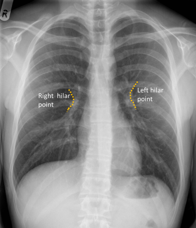
Figure 28: There is cardiomegaly with bilateral prominent hila secondary to enlarged pulmonary arteries in the patient with pulmonary hypertension.
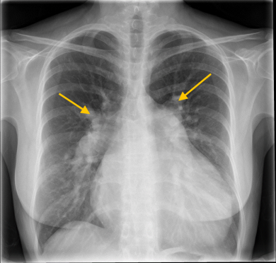
Figure 29: Bilateral enlarged hilar lymphadenopathy in a patient with sarcoidosis.