Top-tip: How to perform an ECGArticle 8 of 13
How to do an ECG
Firstly, familiarise yourself with the machine made available to you. ECG machines come in various levels of complexity from single sheet feeders to strip feeders and some have auto-analyse functions. Basically, there are four limb leads and six pre-cordial chest leads. The chest leads are not colour-coded but the limb leads are. Find the on-off button and make sure you have sticky pads available.
Introduce yourself to the patient, explain what you are going to do and expose the patient’s chest, ankles (remove socks) and wrists (remove watches and jewellery), ensuring privacy. Patients with very hairy chests may need some hair shaving to ensure good contact. Always have a chaperone present for female patients. Stick the sticky electrodes on as follows:
ECG Lead Placement
ECG electrodes comprise of 12 leads which are placed on the limbs and precordium in the following manner:
Image to demonstrate 12 lead ECG electrode placement.
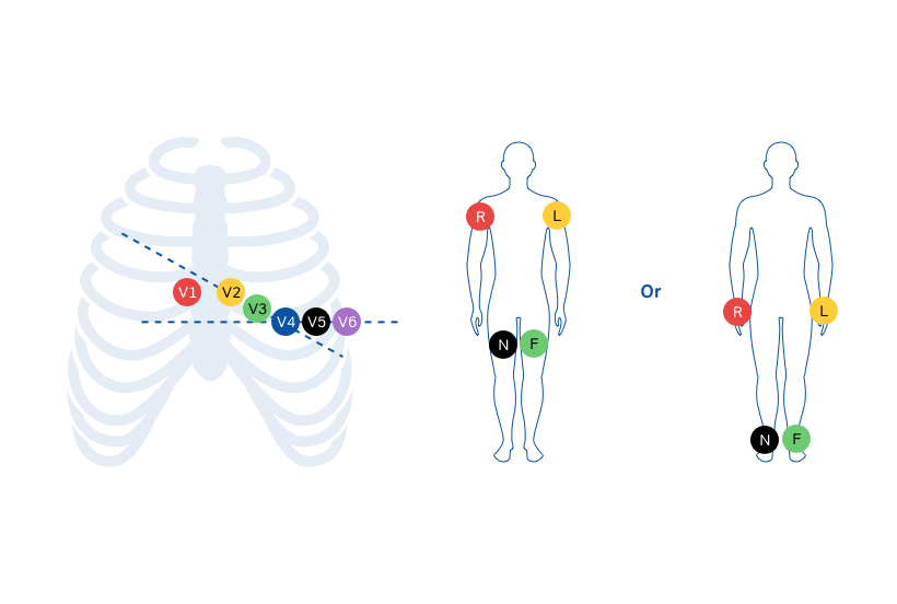
V1: Fourth intercostal space to the right of the sternum.
V2: Fourth intercostal space to the Left of the sternum.
V3: Directly between leads V2 and V4.
V4: Fifth intercostal space at midclavicular line.
V5: Level with V4 at left anterior axillary line.
V6: Level with V5 at left midaxillary line.
(Directly under the midpoint of the armpit)
Limb Electrodes are colour coded Red ( Right Arm), Yellow (Left Arm), Green (Left Leg) abd Black (Right Leg). Stick these on bony prominences on wrist or ankle. ‘Ring Your Green Bell’ (Clockwise from the right arm) is a useful way to remember.
Record the ECG trace and look at it to ensure it is of satisfactory quality before removing the sticky pads from the patient (warn may pull off hairs).